Understanding the differences between the REMS (Radiofrequency Echographic Multi-Spectrometry) scanner and the DEXA (Dual-Energy X-ray Absorptiometry) scanner is crucial for patients concerned about bone health. Both tools are widely used for measuring bone density, but each has its unique features, benefits, and limitations. This article aims to provide patients with a comprehensive understanding of these diagnostic tools, including their processes, results, costs, and which may be better suited to specific needs.
What is a Bone Density Scan?
DEXA and REMS are both non-invasive methods for assessing bone density, but they operate differently. A DEXA scan uses low-dose X-rays to measure bone mineral density (BMD), while REMS utilises ultrasound waves. DEXA is the more established of the two, often considered the gold standard for diagnosing osteoporosis and assessing fracture risk. REMS is a newer technology, gaining attention for its convenient, radiation-free assessment.
Initial symptoms that might necessitate a bone density scan include frequent fractures, height loss, or a stooped posture, often indicative of osteoporosis. Diagnosis through these scans helps doctors determine the extent of bone loss and plan appropriate treatments.
The Scanning Process and Results
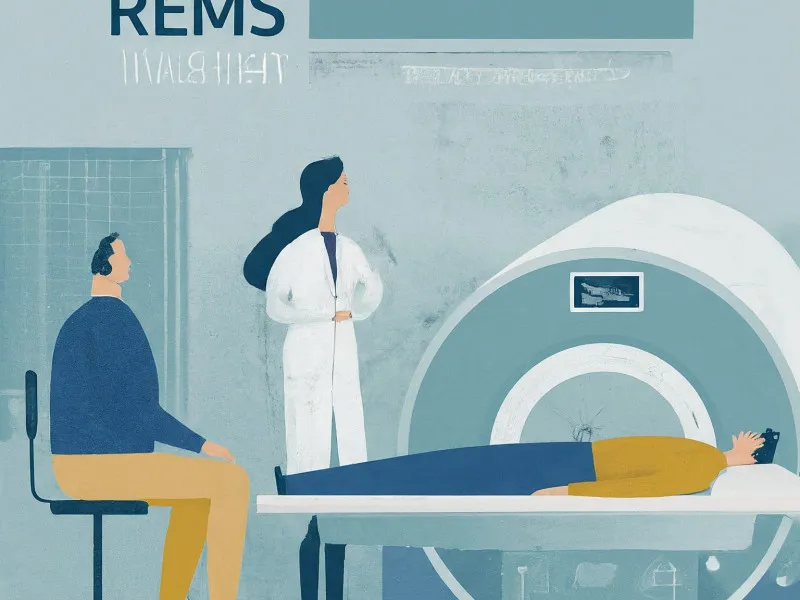
The DEXA scan typically involves lying on an examination table while an X-ray device passes over your body, focusing mainly on the hip and spine since these areas are most prone to fractures. The procedure is quick, usually lasting about 10-20 minutes. REMS, on the other hand, uses an ultrasound probe placed on the surface of the skin, often at the heel bone, and delivers results in a matter of minutes.
Results from a DEXA scan are usually given in T-scores and Z-scores. The T-score compares your bone density to a healthy young adult, while the Z-score compares it to people of your age, sex, and size. A T-score between -1 and -2.5 indicates low bone density, and a score of -2.5 or lower signifies osteoporosis (dexa scan normal range). REMS results are generally considered comparable but can vary based on the device used.
Side Effects and Complications
Both scans are safe, but there are potential side effects and complications. The X-rays used in a DEXA scan expose patients to a minimal amount of radiation. While the radiation dose is very low, some patients may have concerns, particularly if scans are required frequently. REMS does not use radiation, making it a preferable option for those who need to avoid repeated exposure.
If osteoporosis is left untreated, it can lead to severe complications such as fractures, chronic pain, and reduced mobility. Early detection through these scans is crucial to managing the condition effectively and preventing these long-term complications.
Recovery and Rehabilitation
Recovery from the procedures themselves is immediate since they are non-invasive. However, if osteoporosis or low bone density is diagnosed, a comprehensive management plan is necessary, including medication, dietary changes, and specific exercises to improve bone strength. Rehabilitation may involve weight-bearing exercises and balance training to reduce the risk of falls and fractures.
Patients may face barriers to recovery, such as difficulty adhering to prescribed exercise routines or making necessary lifestyle changes. Overcoming these barriers may require support from healthcare providers, physical therapists, and possibly support groups.
Preventing Future Bone Health Issues
Early intervention and regular check-ups are essential. To prevent future issues, patients should maintain a balanced diet rich in calcium and vitamin D, engage in regular physical activity, avoid smoking, and limit alcohol consumption. It’s also important to be aware of the side effects and interactions of any medications taken, particularly those that may affect bone health.
If you experience symptoms like sudden fractures or significant pain, seek medical advice promptly to prevent worsening of the condition.
Frequently Asked Questions (FAQs)
How does a DEXA scan show arthritis?
A DEXA scan primarily measures bone density but can reveal joint health by indicating changes in the bone structure and density around the joints, potentially linking to arthritis (does a dexa scan show arthritis).
Can a DEXA scan show a fracture?
While a DEXA scan is not typically used to diagnose fractures, it can indicate areas of low bone density that are more prone to fractures, helping to identify individuals at risk (can a dexa scan show a fracture).
What is a bad bone density score?
A T-score of -2.5 or below is considered a bad bone density score, indicating osteoporosis. This score signifies a significantly increased risk of fractures (what is a bad bone density score).
What are the normal ranges for DEXA scan results?
A normal DEXA scan result typically has a T-score of -1 and above. Scores between -1 and -2.5 indicate low bone mass (osteopenia), and a score of -2.5 or lower suggests osteoporosis (dexa scan normal range).
What does bone density z-score by age indicate?
The Z-score compares your bone density to that of a typical individual of your age, sex, and size. It’s particularly useful for diagnosing bone density issues in children, young men, and premenopausal women (bone density z-score by age).
What are the advantages of REMS over DEXA?
REMS does not expose patients to radiation and can be performed more frequently without risk. It offers a quick, convenient alternative, especially suitable for individuals who need regular monitoring of their bone health.
In conclusion, both DEXA and REMS provide valuable insights into bone health, but their usage may depend on individual needs and medical advice. Understanding these tools helps patients make informed decisions and take proactive steps towards maintaining strong, healthy bones.